Simple blood test unveils hidden risks of unstable coronary plaques
Researchers have applied a novel assay to indicate impaired HDL function in patients with high-risk coronary plaques
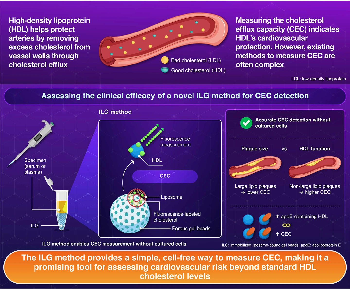
A straightforward blood-based assessment developed at Institute of Science Tokyo, Japan, can help assess how effectively high-density lipoprotein (HDL) remove cholesterol from blood vessel walls, a function known as cholesterol efflux capacity (CEC). The study linked low CEC to a high-risk coronary plaque, supporting the broader clinical use of this method for predicting cardiovascular risk and improving preventive strategies.
Linking HDL Function to Plaque Burden Using Novel ILG Method
Cardiovascular disease is one of the leading causes of death worldwide. Against this background, predicting the risk of coronary artery disease continues to be a major challenge in clinical medicine. High-density lipoprotein (HDL), also known as "good cholesterol," is traditionally associated with protection against coronary plaques. However, conventional HDL measurements do not always reflect how effectively HDL particles function in the body.
One important indicator of HDL function is cholesterol efflux capacity (CEC), which measures the ability of HDL to remove excess cholesterol from cells and transport it to the liver for metabolism. However, conventional methods for measuring CEC are often complex. Addressing this, a research group led by Professor Ryunosuke Ohkawa and Professor Emeritus Minoru Tozuka from the Department of Clinical Bioanalysis and Molecular Biology, Institute of Science Tokyo (Science Tokyo), Japan, previously developed a simple and highly accurate method known as the immobilized liposome-bound gel beads (ILG) method, which uses ILG for CEC determination.
While the method gave promising results, its potential for use in everyday practice was yet to be determined. Now, the researchers, in collaboration with Associate Professor Taishi Yonetsu from the Department of Cardiovascular Medicine, Science Tokyo, together with Mr. Tsunehiro Miyakoshi, a doctoral student in the Department of Clinical Bioanalysis and Molecular Biology, Science Tokyo, analyzed samples from multiple patients to evaluate the clinical utility of the method for detecting the risk of heart disease. The study was made available online in the journal Atherosclerosis on April 2, 2026.
"We analyzed 61 patients who had undergone catheter examinations in the Department of Cardiology," explains the main author, Ohkawa. The researchers measured CEC values from patient samples using the ILG method and compared these with plaque characteristics via optical coherence tomography, an advanced imaging technique capable of visualizing coronary artery plaques in detail.
The results revealed that the patients with high-risk, large lipid-rich plaques had significantly lower CEC values, whereas those with non-large lipid-rich plaques showed higher CEC values. Additionally, higher CEC values were associated with HDL particles containing apolipoprotein E. These findings suggest that lower cholesterol removal capacity may be associated with the development of unstable plaques, which can trigger serious cardiovascular events.
"By simplifying the measurement of CEC, we aimed to make this biomarker more accessible for clinical use," notes Ohkawa.
While large lipid-rich plaques are widely recognized as vulnerable lesions capable of rupturing and triggering acute coronary syndromes, detecting these plaques in living patients without invasive procedures, such as cardiac catheterization, remains challenging. By identifying patients who show reduced HDL function with the simple ILG method, physicians can better predict future cardiovascular risk and guide preventive treatment strategies.
Although CEC has been recognized internationally as a promising biomarker, its widespread use has been limited by the complexity of existing measuring methods. The current ILG method helps overcome this barrier by enabling practical and reliable CEC testing. In future, the researchers hope that the ILG method may contribute to the earlier detection of coronary artery disease risk and more precise monitoring of patients after cardiovascular events.
Reference
- Authors:
- Tsunehiro Miyakoshi1, Yuna Horiuchi1,2, Makoto Araki3, Taishi Yonetsu3, Mei Watanabe4, Takahiro Kameda1,5, Akira Yoshimoto1, Naoya Ichimura4, Shuji Tohda4, Minoru Tozuka1,6, Tetsuo Sasano3, and Ryunosuke Ohkawa1*
- Title:
- Relationship of atherosclerotic lesion by optical coherence tomography with cholesterol efflux capacity by immobilized liposome-bound gel beads method
- Journal:
- Atherosclerosis
- Affiliations:
- 1Department of Clinical Bioanalysis and Molecular Biology, Graduate School of Medical and Dental Sciences, Institute of Science Tokyo, Tokyo, Japan
2Department of Clinical Laboratory Technology, Faculty of Medical Sciences, Juntendo University, Japan
3Department of Cardiovascular Medicine, Graduate School of Medical and Dental Sciences,Institute of Science Tokyo, Japan
4Clinical Laboratory, Institute of Science Tokyo Hospital, Japan
5Department of Clinical Laboratory Science, Faculty of Medical Technology, Teikyo University, Japan
6Life Science Research Center, Nagano Children's Hospital, Japan
Related articles
Further information
Professor Ryunosuke Ohkawa
Department of Clinical Bioanalysis and Molecular Biology, Institute of Science Tokyo, Tokyo, Japan
- ohkawa.alc@tmd.ac.jp
Contact
Public Relations Division, Institute of Science Tokyo
- media@adm.isct.ac.jp
- Tel
- +81-3-5734-2975