How do cells stick to surfaces?
Researchers uncover how the ideal scaffold for cell attachment is created
What the research is about
Cells grown for regenerative medicine or for testing the safety of new drugs must be cultured in artificial environments. However, cells do not simply settle on a culture dish by themselves. Rather than attaching directly to the surface of the dish, cells first anchor themselves to proteins that gather on the surface. These proteins act as a kind of scaffold for the cells. Yet scientists have long been unsure what makes an ideal protein layer for cell attachment.
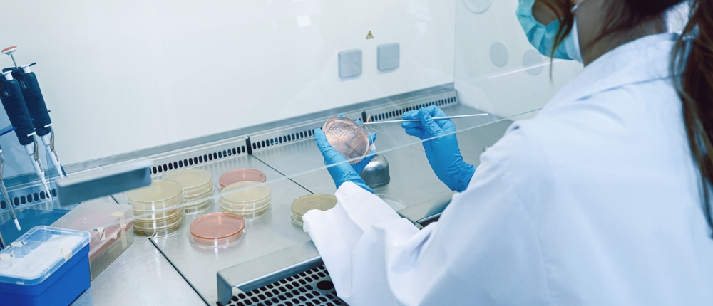
To improve cell culture, various surface treatments are commonly applied to culture dishes. One widely used method is ultraviolet/ozone (UVO) treatment, which uses ultraviolet light and ozone to modify the surface properties of a material. Although it has long been known that UVO treatment improves cell attachment, the reason why remained unclear. Researchers also knew that both insufficient and excessive UVO treatment reduced cell attachment, but the reason for this phenomenon was also unknown.
A research team led by Associate Professor Tomohiro Hayashi of Institute of Science Tokyo (Science Tokyo) set out to solve this mystery by investigating how the protein scaffolds that support cell attachment are formed on UVO-treated surfaces.
Why this matters
Scientists have long known that cells attach to proteins on a surface rather than directly to the material itself. The unanswered question was how UVO treatment creates these protein scaffolds. Instead of focusing on the cells themselves, the research team focused on the scaffolds the cells use. Rather than examining only the surface properties of the material, they tracked which proteins accumulated on the surface and how those proteins were replaced over time.
Previous studies suggested that making a surface more hydrophilic—that is, more water-friendly—would improve cell attachment. However, this idea could not explain why cell attachment reaches its maximum after a short period of UVO treatment and then decreases when the treatment is extended. To address this question, the team examined not only changes in surface chemistry but also the amount, type, and dynamic exchange of proteins on the surface.
Their results revealed that cell attachment was strongest on surfaces treated with UVO for only one to two minutes. After a short treatment, hydrophilic regions formed on the surface, while some of the original hydrophobic (water-repelling) regions remained. This mixed state of hydrophilic and hydrophobic regions turned out to be crucial.
Under these conditions, proteins important for cell attachment accumulated more effectively, creating an ideal scaffold for cells. In contrast, when the treatment was too long, the surface became overly hydrophilic. As a result, the proteins important for cell attachment were less likely to remain on the surface, reducing the availability of suitable scaffolds and ultimately decreasing cell attachment.
The study shows that cell attachment is not determined simply by how hydrophilic a surface is. Instead, it depends on what kind of protein layer forms on that surface. The researchers demonstrated that a slightly heterogeneous surface—not a perfectly uniform one—creates the most effective scaffold for cells.
What’s next
What cells actually interact with is the layer of proteins that forms on the material’s surface. People often assume that making a surface more uniform and cleaner will always improve performance. Surprisingly, the researchers found that cells prefer an environment with a small amount of remaining hydrophobicity—a surface that is not perfectly uniform.
This insight could help guide the development of improved cell culture dishes, artificial organ materials, and regenerative medicine materials. A better understanding of how cells respond to their surroundings may eventually allow scientists to control cell behavior more precisely, opening the door to new biomedical technologies.
Comment from the researcher
Our study showed that the best scaffolds for cells are created when hydrophilic and hydrophobic regions coexist in the right balance. By understanding the relationships among materials, proteins, and cells, we hope to contribute to the development of more advanced biomedical materials.
A major theme of our research is uncovering what happens at interfaces that are invisible to the naked eye. In a previous study, we investigated the interactions between cell membranes and DNA nanomachines (see our related press release article, ‘New tool reveals how DNA nanostructures interact with cell membranes'). This time, we focused on the interface between cells and materials. By understanding these hidden phenomena, we hope to make discoveries that will lead to future medical technologies.
(Tomohiro Hayashi, Associate Professor, School of Materials and Chemical Technology, Institute of Science Tokyo)
Dive deeper
Explore more research in Science for All
Science for All showcases cutting-edge research at Science Tokyo and highlights the ideas, people, and possibilities shaping the future.
Contact
Research Support Service Desk